Reading time : 3 minutes
What is an MRI?
Magnetic Resonance Imaging or MRI is an imaging technique allowing us to obtain pictures of the anatomy and physiological processes of the human body.
This technique uses magnetic fields and computer-generated radio waves to create those images.
As you know, the human body is mainly made up of water. Water molecules are composed of hydrogen and oxygen. Each hydrogen molecule contains an even smaller particle named proton. Those protons are like miniature magnets very sensitive to magnetic fields and this sensitivity is used in magnetic resonance imaging.
Is it painful?
It isn’t painful. The only side effect you could expect is the loud tapping sounds made by the machine while the images are being created.
How to get prepared?
MRI using magnetic properties of the human body to acquire images, scanners contain powerful magnets. For that reason, you will be requested to remove all piercings, medical devices such as cochlear implants, pacemakers, neurostimulator or any other objects susceptible to be attracted.
An IV catheter could be placed to inject medication or contrast.
How does it work?
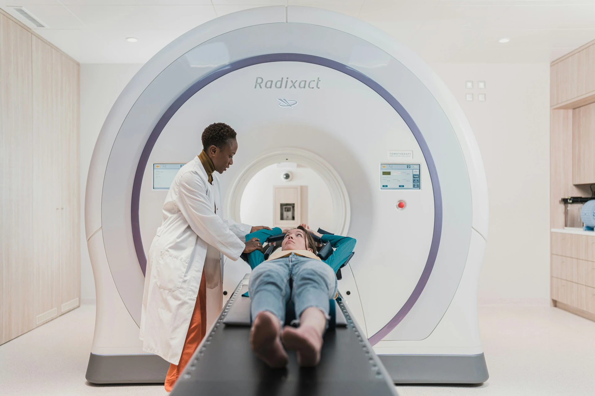
To get the images, you will be asked to enter a large tube scanner. It looks like any other scanner you might have seen in TV shows.
It is very important you stay still during the exam.
Because the pelvis, our area of interest, is located in the middle of the body, you could in most scanners ask to enter feet first rather than head-first, would you be claustrophobic.
The radiographer will be in another room, but you will be able to communicate through an intercom. In most machines, you could also ask for music.
The Pelvis contains the reproductive system but also the urinary and digestive systems. Medication such as spasmolytics diminishing the bowel movement could be injected.
The exam lasts about 15-20 minutes.
What’s the difference with an ultrasound?
MRI allows more precise analyses than abdominal ultrasound. Another main advantage when compared to transvaginal ultrasound performed in non-virgin patients is a broader area of interest.
What should we expect?
In adolescents, endometriosis is rarely advanced. Small lesions, however, are difficult to assess in MRI.
The main purpose of the exam is to evaluate if any other condition could explain your symptoms.
Is there an alternative?
More and more data are collected about salivary tests in endometriosis. You could wonder how salivary tests would help in the diagnosis of endometriosis.
We know endometriosis can be diagnosed in several members of the same family and 50% of the risks of suffering from endometriosis depend on genetics. However, endometriosis is a complex disease depending on numerous factors such as genes, environment, and interactions between both.
MicroRNAs are fragments of RNAs regulating genes. They can be found in blood, urine, and saliva.
Different studies have evaluated RNAs in endometriosis diagnosis. A Yale team have studied RNAs in blood and were able to identify patients with endometriosis. A French team on the other hand have identified 109 mRNAs forming a signature in the saliva of patients with endometriosis. The latter has a 96% sensitivity meaning they could identify 96% of the patients with endometriosis.


