Reading time : 4 minutes
The Critical Role of Transvaginal Ultrasound in Diagnosing and Managing Endometriosis
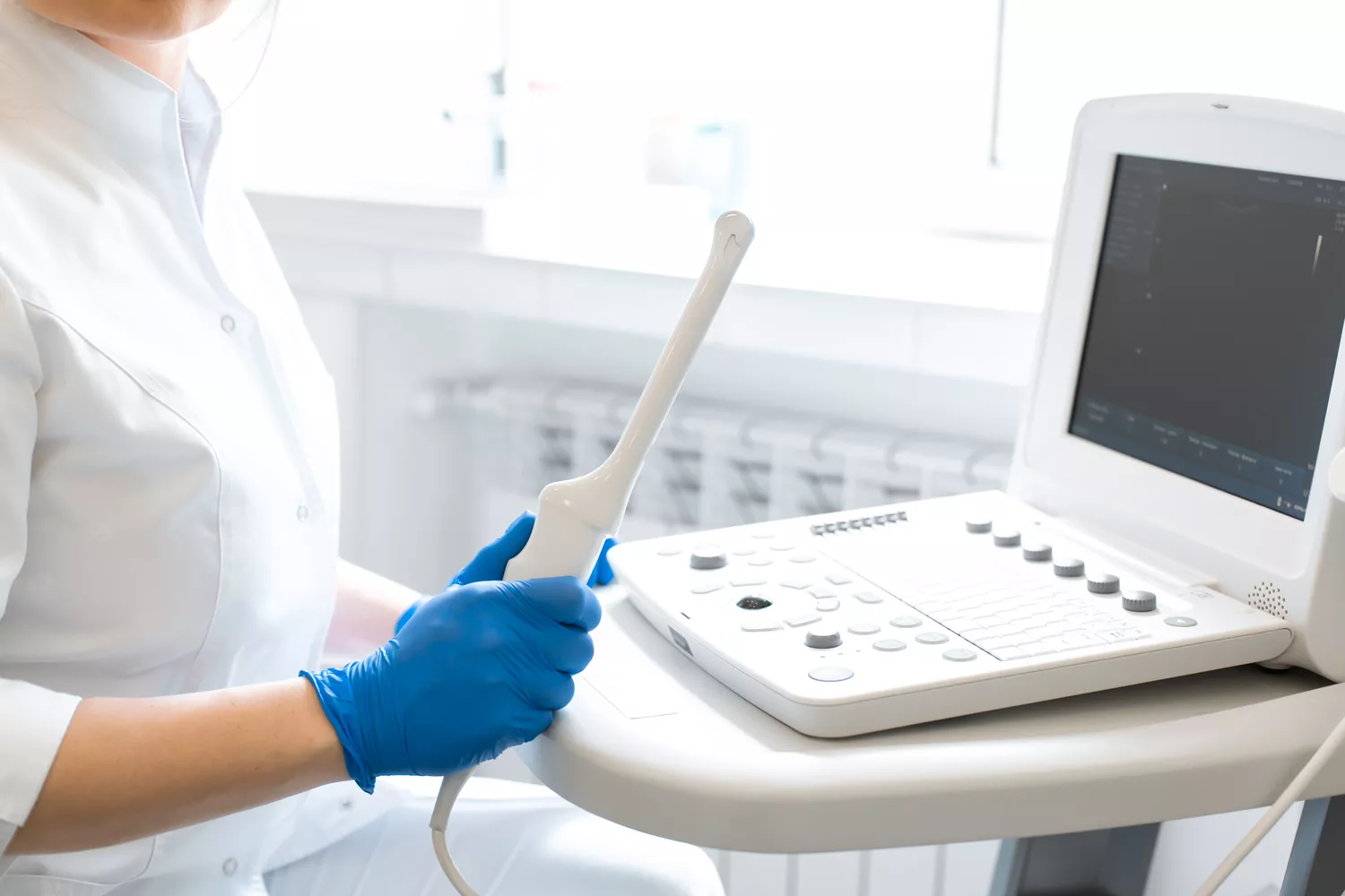
Transvaginal ultrasound (TVS) is widely recognized as the gold standard for diagnosing, describing, and staging endometriosis. As a noninvasive diagnostic technique, TVS offers a crucial tool not only for confirming the presence of endometriosis but also for planning surgical procedures for women who are candidates for laparoscopic treatment. Its ability to provide detailed imaging of the pelvic organs makes it an indispensable part of managing this complex and often debilitating condition.
Understanding the Utility of TVS in Endometriosis
When endometriosis is suspected, TVS plays a pivotal role in explaining the underlying causes of symptoms by identifying the typical ultrasonographic features associated with the disease. Through detailed imaging, TVS allows for the precise mapping of pelvic disease and helps assess its severity, guiding both diagnosis and treatment.
One of the unique aspects of TVS is its use in "pain mapping," a technique where pressure applied through the transvaginal probe helps locate painful nodules. This method is particularly valuable as it provides real-time feedback from the patient, directing the sonographer to areas of concern. Even in cases where ultrasonographic signs are not immediately apparent, endometriosis can still be suspected if there is reduced or absent mobility of pelvic structures—another critical diagnostic clue that TVS can reveal.
Comprehensive Pelvic Examination with TVS
During a TVS examination, the sonographer systematically scans all the main pelvic organs and spaces to identify any abnormalities. This comprehensive evaluation includes:
- Uterus and Adnexa: Key areas for detecting abnormalities related to endometriosis and other gynecological conditions.
- Pouch of Douglas: A frequent site for endometriotic lesions, its assessment is crucial for accurate staging.
- Bladder and Ureters: Important for identifying bladder endometriosis and related complications.
- Rectum and Rectosigmoid Junction: Essential for detecting deep infiltrating endometriosis that may affect bowel function.
- Rectovaginal Septum and Uterosacral Ligaments: Common locations for endometriosis that require careful evaluation.
- Vesicouterine Pouch and Parametria: Areas that need to be thoroughly examined to rule out less common sites of disease.
In addition, the myometrium is meticulously evaluated for signs of adenomyosis, a condition closely related to endometriosis. The presence of direct or indirect ultrasonographic signs of adenomyosis can significantly influence the management plan for the patient.
Standardizing the TVS Examination
To ensure consistency and accuracy in the assessment of endometriosis, the IDEA group has proposed a standardized four-step approach for pelvic ultrasonographic assessment:
- Routine Evaluation of Uterus and Adnexa: The foundational step for detecting common sites of endometriosis.
- Search for Soft Markers: Includes site-specific tenderness and assessing the mobility of pelvic structures.
- Assessment of the Pouch of Douglas: Critical for identifying posterior compartment disease.
- Investigation of the Anterior and Posterior Compartments: Ensures a thorough examination of all potential sites of endometriotic lesions.
When performed according to these steps, TVS achieves an overall sensitivity of 88% and specificity of 79%. This high diagnostic accuracy underscores the importance of TVS as the first-line imaging approach for suspected endometriosis.
Sensitivity and Specificity of TVS
TVS has demonstrated high diagnostic performance across various structures commonly affected by endometriosis:
- Uterosacral Ligaments: Sensitivity of 53% and specificity of 93%.
- Rectovaginal Septum: Sensitivity of 49% and specificity of 98%.
- Bladder Endometriosis: Sensitivity of 62% with perfect specificity (100%).
- Rectosigmoid Deep Infiltrating Endometriosis: Sensitivity of 91% and specificity of 97%.
- Endometrioma: Sensitivity of 73% and specificity of 93%.
These figures highlight the reliability of TVS in diagnosing even complex cases of endometriosis, making it an essential tool for clinicians.
Alternative Approaches for Adolescents
While TVS is the preferred first-line imaging technique, it may not be well-tolerated by all patients, particularly adolescents who may experience discomfort or who are virginal. In such cases, transrectal or transabdominal approaches can be proposed as alternatives. Transabdominal scanning, in particular, can enhance the accuracy of the examination, especially when dealing with extrapelvic disease or extensive findings.
Conclusion
Transvaginal ultrasound is a cornerstone in the diagnosis and management of endometriosis, offering unparalleled insights into the extent and severity of the disease. Its dynamic nature and the potential for real-time patient interaction during the examination make it superior to many other imaging techniques. By following standardized assessment protocols, healthcare providers can ensure high diagnostic accuracy, ultimately leading to better outcomes for women suffering from endometriosis.


